
What are meibomian glands?
Meibomian glands are sebacous (oil) glands located in the upper and lower eyelids. There are more in the upper lid than the lower lid, and they are also larger in the upper lid (1). The glands sit vertically in the eyelids, and their openings are right behind the lash line. The job of the meibomian glands is to make and secrete oil. These secretions make up one of the 3 layers of the tear film, the lipid layer. Blinking helps spread the secretions on the surface of the eye, keeping the tear film stable and preventing it from evaporating too quickly. The function of the glands is regulated by hormones such as androgens, estrogens, and progestins.

Illustration of the meibomian glands, showing how they are oriented vertically in the upper and lower lid // via: All About Vision
What is Meibomian Gland Dysfunction?
Meibomian gland dysfunction, or MGD, is not the most clearly defined condition. Here’s the technical definition:
“Meibomian gland dysfunction (MGD) is a chronic, diffuse abnormality of the meibomian glands, commonly characterized by terminal duct obstruction and/or qualitative/quantitative changes in the glandular secretion. It may result in alteration of the tear film, symptoms of eye irritation, clinically apparent inflammation, and ocular surface disease.” (2)
Translation? MGD is when the meibomian glands do not work properly. The dysfunction is typically either a result of a blockage in the gland or an abnormality in the oil being secreted. A blockage may present as a clear or opaque dome at the opening of the glands (see photo below). MGD can also involve changes in the quality and quantity of secretions. When pressure is applied to normal glands, they should secrete a small amount of clear oil. In the case of MGD, the secretion is thicker and more yellow-white in color. I liken it to when you leave a tube of toothpaste open for a while, and you get some crusty, solidified gunk at the opening that you have to squeeze out to get to the usable toothpaste. When the oil glands do not function properly, that leaves the tearfilm deficient of oils, thus leaving it unstable and leading to dry eye symptoms. If left untreated, this chronic condition can result in progressive meibomian gland destruction.
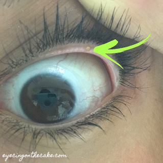
The yellow dome seen here is a visibly capped gland
What are the signs and symptoms of MGD?
Some common symptoms include dryness, a sandy/gritty feeling, a burning sensation, eye and lid irritation, contact lens intolerance, eyelids sticking together in the morning, and even blurry vision. These symptoms can occur as a result of other conditions, therefore a thorough examination of the eyes, eyelids, and tear film is warranted to help identify the source of the problem. Common associated signs of MGD are thickened eyelid margins, frothy tears, and a low tear break-up time (the time it takes for the tear film to break up due to evaporation). In fact, dysfunction of the meibomian glands is one of the main causes of evaporative dry eye disease. Not only does it cause dryness, but insufficient lipids may cause increased bacterial growth on the lid margins, which can cause a number of secondary lid issues (2).

The top: an eyelid with normal meibomian glands (note that the glands are straight, extending down the eyelid)
The bottom: an eyelid with meibomian gland dysfunction (note that the glands are curlier and shorter than the top image)
What is MGD associated with?
- Age: MGD increases in prevalence with age (3).
- Digital device use: We blink less often and less completely when on digital devices, which effects the secretion of oil from the eyelids.
- Contact lens wear: Studies suggest a decrease in functional meibomian glands with contact lens wear (4, 5).
- Ethnicity: MGD appears to have a much higher prevalence among Asian populations (2).
- Systemic factors: Examples include menopause, rosacea, Sjogren’s syndrome.
- Medications: Certain medications, like Accutane, as well as preservatives used in eye drops, like BAK, have been linked to increased incidence of MGD (6).
How is MGD treated?
There are various treatment options available, depending on the severity and associated symptoms/signs. Here is a list of treatments I typically recommend for MGD, going from least complex (for milder cases) to most complex.
1. Blink!
Blinking stimulates the secretion of meibum, and helps spread it across the surface of the eye. Studies show you blink less when reading, and you blink 60% less when at the computer (7). So take frequent breaks, and make a conscious effort to blink completely during those breaks.

Image via Optometric Management
2. Warm compresses with lid massage
Taking a page out of a colleague’s book, it’s the flossing of the eye world– it’s something you SHOULD do daily to prevent disease, but not a lot of people do it religiously.
- Use a warm compress (commercially-available ones like Bruder masks or Tranquileyes, or make your own using a boiled egg or warmed dry rice wrapped in a clean cloth- something that stays warms for a few minutes) and rest it over your eyelids with your eyes closed. Do so for about 10 minutes. Gently massage your eyelids, rolling your fingers vertically down your upper lid and up your lower lid (towards your lashes). This helps get the oils flowing, and removes any obstruction within the glands as well as any solidified gunk at the opening of the glands.
- I recommend doing this twice daily when symptoms are present, or when MGD is first diagnosed. Beyond that, once daily is great for maintenance.
- There are now in-office procedures that have the same goal of melting meibum and removing gland obstruction, and they do so more effectively. Lipiflow and iLux are examples. These devices heat the internal surface of the lids and simultaneously applies pressure to the external lid to express the glands, all in a few minutes.
3. Lid cleanser
Ideally, you would follow up your warm compress and lid massage by cleaning the eyelids and eyelashes. Using a lid cleanser, gently scrub along the lash line to remove debris, makeup, and bacteria that may clog or infect the oil glands. A cleanser containing diluted tea tree oil is particularly beneficial if Demodex is present. Hypochlorous acid (HCA) spray is also a good option. HCA helps control bacterial overgrowth and degrades disruptive enzymes that play a role in inflammation.
4. Omega-3 fatty acids
Omega-3s help improve the quality of the oil produced by the meibomian glands, and they have anti-inflammatory effects. A great source of omega-3s is fatty fish, like salmon and tuna. Another option is fish or flax seed oil supplements. It’s always a good idea to consult with your doctor before starting any supplement.
5. Artificial tears
Artificial tears can also help beef up the contents of the tear film. Lipid-based artificial tears are best in the case of MGD because they help replenish the lipid layer of the tear film. Some lipid-based drops: Systane Complete, Soothe XP, Retaine MGD, and Refresh Mega-3. Preservative-free drops are ideal whenever possible.
6. Prescription medications
Depending on the other presenting conditions, some patients with MGD may require the use of antibiotics. Topical azithromycin or low-dose, long-term use of oral doxycycline are thought to alter the eyelid bacteria and also provide anti-inflammatory effects (8, 9). Still others with severe MGD may need to have the inflammation controlled through the short-term use of a topical steroid drop or the long-term use of a dry-eye drop.
| CLIFFSNOTES: Meibomian gland dysfunction (MGD) is a common condition in which the oil glands in your eyelids do not function properly. MGD can alter the makeup of the tear film, leading to eye irritation, inflammation, and dry eye. There are many different ways to treat MGD and dry eye, so it is best to start with a thorough examination at your eye doctor’s office to gather all the data, identify risk factors, and formulate a treatment plan that is specific to you. So see your eye doctor STAT to nip it in the bud! |